Retinopathy of Prematurity (ROP) is a potentially serious eye condition that affects premature infants. It is characterized by abnormal blood vessel growth in the retina, the light-sensitive tissue at the back of the eye. Understanding the pathophysiology of ROP is crucial in comprehending the underlying mechanisms and factors contributing to the development and progression of this condition.
Retinopathy of Prematurity
Retinopathy of Prematurity (ROP) is a potentially sight-threatening condition that primarily affects premature infants. It is characterized by abnormal blood vessel development in the retina, the light-sensitive tissue at the back of the eye.
Causes of Retinopathy of Prematurity
The exact cause of ROP is not fully understood. However, it is believed to occur due to the following factors:
- Prematurity: ROP primarily affects premature infants who are born before 31 weeks of gestation or with a birth weight below 1500 grams (3.3 pounds). The blood vessels in the retina of premature infants may not have fully developed making them vulnerable to abnormal growth.
- Oxygen therapy: Premature infants often require supplemental oxygen to assist with breathing. However, high levels of oxygen or fluctuations in oxygen levels can disrupt the normal development of blood vessels in the retina leading to ROP.
- Other risk factors: Certain factors can increase the risk of developing ROP including low birth weight, respiratory distress, blood transfusions, anemia and infections.
Symptoms of Retinopathy of Prematurity
In the early stages, ROP may not cause any noticeable signs or symptoms. However, as the condition progresses, the following symptoms may become apparent:
- Abnormal eye appearance: The eyes may appear unusual, with dilated pupils or whitish color in the center of the eye (pupil).
- Strabismus: Also known as crossed eyes, strabismus is a condition in which the eyes do not align properly.
- Nystagmus: Nystagmus refers to involuntary eye movements which may be rapid and jerky.
- Poor visual tracking: Infants with ROP may have difficulty visually tracking objects or following movements.
It is important to note that only a comprehensive eye examination by an ophthalmologist can accurately diagnose ROP.
Classification and Stages of Retinopathy of Prematurity
ROP is classified into different stages based on the severity of the condition and the extent of abnormal blood vessel growth. The stages include:
- Stage 1: Mildly abnormal blood vessel growth with a thin white line appearing in the retina.
- Stage 2: Moderately abnormal blood vessel growth characterized by a ridge or fold in the retina.
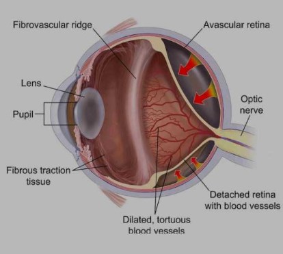
- Stage 3: Severely abnormal blood vessel growth where the new blood vessels extend into the clear, gel-like substance (vitreous) in the center of the eye.
- Stage 4: Partial retinal detachment with the retina pulling away from the back of the eye.
- Stage 5: Total retinal detachment where the retina is completely detached, leading to potential vision loss or blindness.
Pathophysiological Process
The pathophysiology of ROP involves several complex processes that occur within the developing retina of premature infants. It can be broadly categorized into two phases:
Phase 1: The Phase of Vasoconstriction
During the early phase of ROP which typically occurs within the first few weeks after birth, the premature infant’s retina is exposed to an oxygen-rich environment outside the womb. This sudden change triggers a series of events:
- Oxygen-induced vasoconstriction: The elevated oxygen levels cause vasoconstriction leading to reduced blood flow through the developing retinal blood vessels.
- Retinal hypoxia: The restricted blood flow results in inadequate oxygen supply (retinal hypoxia) to the developing retina.
- Release of angiogenic factors: In response to hypoxia the retina releases various angiogenic factors including vascular endothelial growth factor (VEGF).
- Incomplete vascularization: The abnormal levels of angiogenic factors disrupt the normal retinal vascular development leading to incomplete retinal vascularization.
Phase 2: The Phase of Neovascularization
As the disease progresses, the phase of neovascularization begins, characterized by the following events:
- Neovascularization: The release of angiogenic factors triggers the growth of abnormal blood vessels (neovascularization) from the retina into the vitreous, the gel-like substance filling the eye.
- Fibrous tissue formation: The neovascularization process can result in the formation of fibrous tissue which can contract and lead to retinal detachment.
- Complications: Retinal detachment can cause severe vision impairment or blindness if not promptly treated.
Risk Factors
Several risk factors contribute to the development and progression of ROP:
- Prematurity: The degree of prematurity plays a significant role in the development of ROP. The more premature the infant, the higher the risk.
- Low birth weight: Infants with low birth weight are more susceptible to ROP.
- Oxygen therapy: The administration of supplemental oxygen to premature infants can disrupt the delicate oxygen balance in the developing retina, contributing to ROP.
- Infection and sepsis: Infections and sepsis can further increase the risk of ROP in premature infants.
- Anemia: Insufficient red blood cells or low hemoglobin levels (anemia) can exacerbate retinal hypoxia and contribute to ROP development.
Prevention and Management
Prevention and appropriate management of ROP are crucial to minimize the risk of vision impairment or blindness. The following strategies are commonly employed:
- Oxygen therapy optimization: Close monitoring and careful regulation of oxygen levels in premature infants to maintain a balance between adequate oxygenation and avoiding excessive oxygen exposure.
- Early screening and diagnosis: Regular eye examinations by an ophthalmologist to detect and monitor the progression of ROP.
- Laser therapy: In cases where ROP reaches an advanced stage laser treatment can be performed to destroy abnormal blood vessels and prevent retinal detachment.
- Anti-VEGF therapy: In some instances anti-VEGF medications may be administered to inhibit abnormal blood vessel growth.
- Surgical intervention: Surgical procedures such as vitrectomy or retinal detachment repair may be necessary for advanced cases of ROP.
Retinopathy of Prematurity (ROP) is a complex condition involving abnormal blood vessel growth in the retina of premature infants. Understanding the pathophysiology of ROP is essential in comprehending the underlying processes that contribute to its development and progression. Identifying risk factors, early detection and appropriate management are crucial in minimizing the potential long-term visual complications associated with ROP.